Bio 113 Portfolio
  • Home
  • Prion
    • Life Cycles
    • Evolutionary History
    • Kuru
    • Sources
  • Virus
    • Life Cycles
    • Evolutionary History
    • Rabies
    • Sources
  • Bacteria
    • Life Cycles
    • Evolutionary History
    • Pseudomonas syringae
    • Borrelia burgdorferi
    • Sources
  • Archaea
    • Life Cycles
    • Evolutionary History
    • Sulpholobus acidocaldarius
    • Sources
  • Protista
    • Life Cycles
    • Evolutionary History
    • Giardia lamblia
    • Phytophthora infestans
    • Hexacontium enthacanthum
    • Corallina officinalis
    • Discostelium discoideum
    • Sources
  • Plantae
    • Life Cycles
    • Evolutionary History
    • Dicranum polysetum
    • Adiantum aleuticum
    • Ginkgo biloba
    • Pyrus communis
    • Magnolia virginiana
    • Sources
  • Fungi
    • Life Cycles
    • Evolutionary History
    • Lentinula edodes
    • Neurospora crassa
    • Gigaspora gigantea
    • Rhizopus stolonifer
    • Batrachochytrium dendrobatidis
    • Sources
  • Animalia
    • Life Cycles
    • Evolutionary History
    • Euplectella aspergillum
    • Lingula anatina
    • Priapulus caudatus
    • Mellita quinquiesperforata
    • Tachyglossus aculeatus
    • Sources

Distinguishing Characteristics

bacterial cell (25)

Cell Structure
  • Unicellular:  composed of a single prokaryotic cell (1)
  • Cell diameter ranges .5-5 um (1)
  • No membrane bound organelles even though the cell is very well organized (1)
  • Presence of 70S ribosomal RNA (20)
Cell Accessories
Accessory(1)
  1. Cell wall
  2. Plasma membrane
  3. Capsule/ Slime layer
  4. Pili/fimbriae
  5. Ribosomes
  6. Nucleoid region
  7. Bacterial flagella
  8. Plasmids (not pictured)
Structure(1)
  1. Rigid wall that contains peptidoglycan
  2. Phospholipid bilayer
  3. Sticky layer of polysaccharide or protein; if dense called capsule, called slime layer if less well organized
  4. Hairlike appendages
  5. Made of ribosomal RNA and protein
  6. Contains single copy of chromosome
  7. Rotary structure driven from a motor at the base, not covered by plasma membrane. Composed of protein flagellin
  8. Smaller rings of separately replicating DNA


Function(1)
  1. Maintains cell shape, protects the cell, prevents from bursting in hypotonic environment
  2. Contains cell contents
  3. Enables bacteria to adhere to substrate or to other bacteria, some prevent dehydration or shield attack from the host's immune system
  4. Pili:  pull two cells together prior to DNA transfer from one cell to another, fimbriae:  stick bacteria to their substrate or to one another
  5. Protein factory of the cell
  6. Contains the DNA
  7. Provides movement
  8. Carry and spread genes to other bacteria, can be horizontally transferred between cells
Types
Shapes:
Coccus- Spherical or oval arrangements, based on planes of division
.5-1um diameter
(3)

pic is (8)
Picture

Bacillus- Rod shaped, all divide in one plane
.5-1um long
(3)

pic is (9)

Picture

Spiral- 1um to 100um long


(3)

pic is (10)
Picture
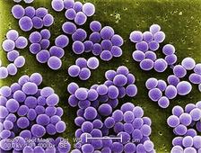
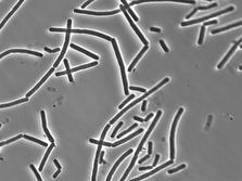
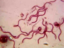
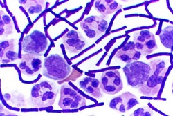
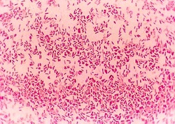
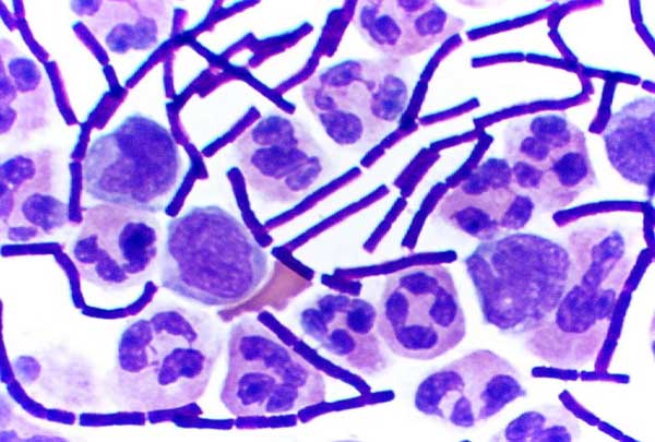
Gram Stain:  bacterial staining technique used to distinguish gram positive and gram negative bacteria by the differences in their cell walls (11)
Picture
pic (12)
Picture
Gram positive: Simpler walls with relatively large amount of peptidoglycan (1)

Stains purple (1)

pic (13)

Picture
Gram negative: Less peptidoglycan, structurally more complex, with an outer membrane that contains liposaccharides (1)

stains pink (1)

pic (14)

Modes of Nutrition (1)
  • photoautotrophy- use photosynthesis to drive synthesis of organic compounds from CO2 or other inorganic carbon compounds
  • chemoautotrophy- oxidize inorganic compounds to obtain carbon
  • photoheterotrophy- obtain energy from the sun, but must get carbon in organic form
  • chemoheterotrophy- consume organic molecules for energy AND carbon
Powered by Create your own unique website with customizable templates.